In the field of vascular diagnostics, angiography has emerged as a critical imaging tool. It enables medical practitioners to visualize blood vessels in detail and effectively diagnose various cardiovascular, neurological, and peripheral conditions. By administering contrast dye into veins or arteries, angiography facilitates real time evaluation of vessel irregularities such as narrowing, blockages, aneurysms, and other anomalies.
Whether you're managing a radiology department, assessing the value of imaging technologies, or working toward improved patient care, understanding the latest developments in angiographic methods is vital.
What Exactly Is Angiography?
Angiography is a diagnostic imaging technique that captures highly detailed images of the body’s vascular system. It involves injecting a contrast agent into the circulatory system to highlight veins and arteries during imaging, typically via X-ray technology. Essentially, an angiogram acts as a moving X-ray that helps observe blood flow through specific body areas.
This method allows clinicians to detect issues like clots, arterial narrowing, or aneurysms. These findings often guide treatments such as embolization, surgical planning, or stent placement. Although "arteriography" traditionally referred to imaging of arteries specifically, the term "angiography" is now more widely used to include both arterial and venous studies. However, unless specified (like in venography), angiography usually pertains to arterial imaging.
Protect Patient Data. Go Cloud. Try Our PACS Absolutely Free
Try Now !Modern procedures often utilize digital subtraction angiography (DSA), which removes non vascular background structures from images, producing clear, high definition visuals of the blood vessels. This accuracy is essential in high stakes situations, such as evaluating heart vessels after a suspected heart attack or inspecting brain circulation following a stroke.
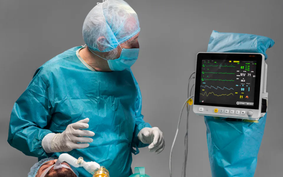
Demonstration of the Angiographic Workflow
Want to see what angiography looks like in a real world application?
With Scriptoware’s integrated DICOM Viewer, users can access angiographic images directly via web or mobile platforms. The DICOM files are stored securely in the Scriptoware cloud PACS, enabling easy and efficient image viewing within a protected workspace.
Different Types of Angiography and Their Medical Roles
Various angiography procedures exist to examine different parts of the body, and the selection typically depends on the patient’s condition and clinical requirements:
- Cerebral Angiography:Used to assess the brain’s vascular network. It helps in diagnosing conditions such as arteriovenous malformations (AVMs), brain aneurysms, and strokes by pinpointing vessel abnormalities that could lead to ischemia or hemorrhage.
- Coronary Angiography:This technique evaluates the coronary arteries that supply the heart. Usually performed in catheterization labs, it helps detect blockages or plaque buildup, offering immediate treatment options such as balloon angioplasty or stenting during the same session.
- Pulmonary Angiography:Primarily performed to locate blood clots in the lungs (pulmonary embolism). Although CT pulmonary angiograms are widely used today, traditional catheter based angiography is still valuable in complex or urgent diagnostic scenarios.
- Peripheral Angiography:Targets the arteries and veins in the limbs. It is especially useful for diagnosing peripheral artery disease (PAD), a condition marked by reduced blood flow to the legs and feet. The results can support decisions involving stent placement, bypass procedures, or other vascular interventions.
These angiographic methods produce detailed, high resolution images that assist physicians in identifying even minor disruptions in blood flow or vessel structure. As such, angiography remains an essential diagnostic and interventional tool across many areas of medicine, particularly in emergency settings.
Angiography Compared to Other Imaging Techniques
When determining the most appropriate imaging technique, healthcare providers consider a range of factors, such as image clarity, invasiveness, patient condition, and the tools available at their facility.
Magnetic Resonance Angiography (MRA) offers a non invasive option that does not involve ionizing radiation. This makes it safer for patients who are more vulnerable or sensitive to radiation. In contrast, traditional catheter based angiography is invasive but offers real time imaging and the potential for immediate therapeutic actions. This is especially valuable in emergency cases.
Terms like “arteriogram” and “angiogram” are often used interchangeably. However, there is a subtle distinction. “Arteriogram” specifically focuses on arterial imaging, while “angiogram” can include both arterial and venous imaging.
CT angiography (CTA) is another diagnostic tool widely used for its speed and ability to generate detailed 3D images. However, it does expose patients to radiation and does not allow for simultaneous treatment like catheter based methods. Doppler ultrasound, on the other hand, is radiation free and non invasive. It is often used to evaluate blood flow, but its resolution can be limited in deep or complex vascular regions, such as those in the heart or brain.
No imaging solution is universally superior. The best option depends on the urgency of the situation, the patient's health status, and the vascular region under investigation.
Angiography’s Role in Diagnosing Vascular Conditions
One of the key strengths of angiography is its ability to offer live, detailed views of blood flow. This helps healthcare professionals identify abnormalities in the vascular system with high accuracy.
Doctors often rely on angiograms to measure the extent of artery narrowing, locate aneurysms, or find the origin of internal bleeding, particularly in trauma cases. Beyond diagnostics, angiography can also be therapeutic. For example, if an issue like a ruptured vessel or a blocked artery is found, immediate treatment such as placing a coil, sealing a vessel, or inserting a stent can often be carried out during the same session.
This combination of diagnosis and treatment in a single procedure is a major advantage. A patient undergoing coronary angiography may receive a stent or undergo angioplasty on the spot if a blockage is detected.
That said, angiography has its limitations. As an invasive procedure, it involves introducing contrast dye into the bloodstream. This may pose risks for patients with kidney issues or dye allergies. Additionally, it tends to be more expensive than non invasive options. These factors make careful patient selection important when alternatives exist.
Explore and evaluate medical images using Scriptoware’s cutting edge DICOM viewer technology.
Try Now !The Financial Aspect of Angiography
The price of an angiography procedure can vary significantly. This depends on several factors, such as the anatomical area being examined, the complexity of the case, facility type, and local healthcare policies.
For example, a cerebral angiogram usually costs more than a peripheral one due to the precision and risk involved. Specialized tools, highly trained staff, and advanced imaging systems also contribute to higher overall costs.
In many healthcare systems, insurance plays a major role in making angiography financially accessible. If the procedure is deemed medically necessary, coverage may be partial or full, depending on the patient’s plan.
In the United States, the price may vary between outpatient imaging centers and hospital settings, even if the services provided are similar. In countries with publicly funded healthcare systems, such as Canada or several European nations, the patient’s out of pocket costs may be minimal. However, this can sometimes result in longer wait times.
To reduce costs without compromising patient care, hospitals and clinics are adopting efficiency measures. These include optimizing appointment scheduling, carefully reusing certain single use materials when safe, and selecting cost effective contrast agents.
What Happens During an Angiography Procedure?
Knowing what to expect before, during, and after angiography can help patients feel more prepared and informed. The duration of the procedure can range from 30 minutes to several hours, depending on whether therapeutic actions are taken during the session.
The process begins with a thorough evaluation of the patient’s medical background. Kidney function is assessed due to the use of contrast dye, and the risks of the procedure are discussed with the patient.
On the day of the exam, the patient is prepped in a specialized imaging suite. Typically, local anesthesia is applied, and a sedative may be given to keep the patient comfortable. A thin catheter is inserted into a blood vessel, usually in the wrist or groin, and navigated to the targeted area. Contrast dye is then introduced to make the vessels visible on the imaging screen.
Once imaging is complete, or after any necessary interventions, the catheter is withdrawn. Firm pressure is applied to the insertion site to prevent bleeding. Afterward, patients are monitored for several hours, particularly if sedation or more invasive steps were involved.
post procedure, it is generally recommended that patients avoid heavy physical activity for a day or two. Written aftercare instructions are provided to minimize the risk of complications such as bleeding, infection, or re-narrowing of the vessels.
Angiography Evolving Technology, Safety, and Its Role in Modern Healthcare
Key Risks and Safety Measures
While angiography remains vital in diagnostic imaging, it carries certain inherent risks. Patients may experience bleeding at the catheter site, allergic responses to contrast material, or, though rarely, damage to blood vessels. For individuals with compromised kidney function, the use of contrast agents could potentially worsen renal performance. There is also a minor risk of stroke or cardiac events if clots or plaque dislodge during the process.
To manage these risks, healthcare providers follow strict safety protocols. These include evaluating kidney health beforehand, using the smallest necessary dose of contrast dye, and ensuring trained specialists perform the procedure. Medical institutions also reduce complications through procedural checklists and regular staff training. This is especially important for older or high risk patients.
Advancements in Angiographic Imaging
The field of angiography is undergoing significant change thanks to progress in technology and computing. One major advancement is the use of artificial intelligence to assist in analyzing angiograms. AI systems can detect abnormalities quickly and help assess risk, making diagnostics faster and more precise.
Another innovation is hybrid imaging, which merges data from CT or MRI scans with live catheter based imaging. This integration enhances visualization and helps guide more complex interventions. Alternatives such as carbon dioxide (CO2) angiography and intravascular ultrasound are becoming popular as well, especially for patients sensitive to Iodine based contrast dyes.
Angiography’s Shifting Place in Healthcare
With increasing pressure to deliver quality care efficiently, angiography remains a cornerstone in diagnostic medicine. Telehealth technologies now enable specialists to review angiograms remotely, speeding up access to expert interpretations. Portable angiography units are also proving useful in emergency settings and rural areas with limited resources.
To control costs, healthcare providers are adopting strategies like shared use of imaging devices and bulk purchasing of medical supplies. As insurance coverage expands for high tech imaging, access is growing. The combined impact of technological innovation and smarter resource use will help define angiography’s role going forward.
Frequently Asked Questions
How is angiography performed?
The process starts with reviewing a patient’s medical background and conducting tests such as kidney function checks. A local anesthetic is applied to the wrist or groin, where a catheter is inserted and guided via X-ray to the target area. Contrast dye is then injected to highlight blood vessels for imaging. Afterward, the catheter is removed, pressure is applied, and the patient is monitored to prevent complications.
Does angiography hurt?
Angiography usually involves minimal discomfort. Patients often feel only slight pressure or a brief warm sensation when the dye is injected. Local anesthesia prevents most pain, and updated techniques ensure that the experience is as comfortable as possible.
Final Thoughts
Despite newer, less invasive options such as MR angiography and Doppler ultrasound, traditional angiography still offers unmatched clarity and real time treatment capabilities. Its role in diagnosing and managing complex vascular issues remains essential. By focusing on safety, cost efficiency, and continued innovation, healthcare providers can ensure angiography remains a crucial diagnostic tool well into the future.
Share your imaging investigations (PET CT, MRI, CT) with your doctor for free
Sign up with Scriptoware