What Does Medical Imaging Mean?
Medical imaging refers to the diverse set of techniques used to visualize internal body structures for medical diagnosis and treatment management. These imaging technologies have become indispensable for detecting illnesses, monitoring progression, and guiding therapy all without invasive procedures. The rise of non invasive imaging tools has transformed healthcare by offering critical insights into anatomy and pathological conditions.
Common Technologies Employed in Medical Imaging
Various advanced technologies capture detailed images from inside the body, each with unique strengths:
- X-ray Imaging: One of the oldest imaging techniques, this method uses radiation primarily to visualize bones and skeletal features.
- Computed Tomography (CT): CT scanning creates detailed cross sectional images by combining multiple X-rays taken from different directions, providing a layered view that surpasses traditional X-ray clarity.
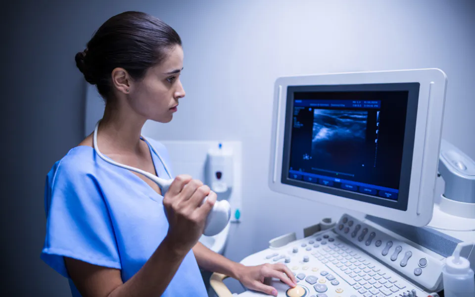
- Ultrasound: This modality utilizes sound waves at high frequencies to produce images of organs and soft tissues. It is especially useful in fields such as prenatal care, cardiac assessments, and musculoskeletal studies.
- Positron Emission Tomography (PET): By injecting a radioactive tracer, PET scans highlight the metabolic activity in tissues, making it valuable for identifying cancer and other metabolic disorders.
- Nuclear Medicine: This field uses trace amounts of radioactive substances for both diagnostic imaging and treatment. Single Photon Emission Computed Tomography (SPECT) is a key technique within this category.
Understanding Magnetic Resonance Imaging (MRI)
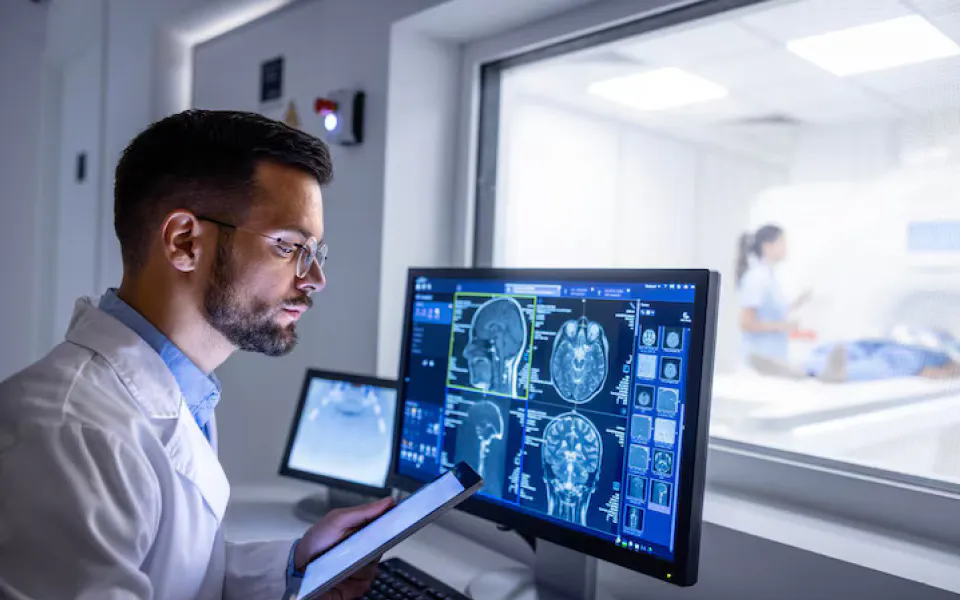
Magnetic Resonance Imaging, commonly known as MRI, creates detailed internal images by using strong magnetic fields combined with radiofrequency waves. This imaging technique excels at producing clear visuals of the brain, spinal cord, and joints.
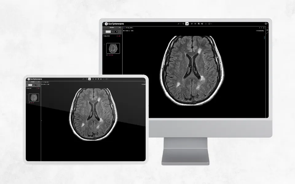
With Scriptoware’s cloud based system, you can securely access your MRI scans anytime using their online DICOM viewer, available on both web browsers and mobile devices. All imaging files are safely stored in Scriptoware’s PACS and cloud workspace for convenient retrieval.
The Working Principles Behind Medical Imaging
Different medical imaging methods rely on unique technologies and physical concepts to visualize the body's interior. Below is a summary of how these main imaging modalities function:
- X-ray Radiography: In this approach, X-rays pass through the body and are absorbed by tissues to different degrees. Bones, being dense, absorb the most and appear as bright white areas, while softer tissues show up in varying gray shades.
- Computed Tomography (CT): CT scanners rotate an X-ray source around the patient, capturing multiple images from various perspectives. These images are then digitally combined to form detailed cross sectional views of internal body parts.
- Magnetic Resonance Imaging (MRI): The patient lies within a strong magnetic field while radiofrequency pulses temporarily realign hydrogen atoms in the body. Signals emitted as the atoms return to their original positions are processed to produce high resolution images.
- Ultrasound: This technique employs a handheld transducer to send high frequency sound waves into the body. Reflected echoes are converted into live images of organs and soft tissue.
- PET and SPECT Imaging: These nuclear medicine techniques involve injecting radioactive tracers into the bloodstream. Gamma rays emitted by the tracers are detected by specialized cameras, enabling visualization of metabolic activity and organ function.
Components of Medical Imaging Technology
The term medical imaging technology involves the combined use of specialized devices and software aimed at visualizing the internal anatomy of the body. It includes physical equipment like MRI machines as well as digital platforms for processing and analyzing images. Thanks to recent innovations, scanning speeds have increased, image quality has improved, and new imaging methods have emerged, which expand diagnostic and treatment possibilities.
By offering detailed views of internal body structures, these technologies are vital for clinical decision making. Although some imaging methods expose patients to radiation, the benefits to health generally surpass the potential risks. Ongoing progress in both imaging hardware and software continues to enhance diagnostic precision and improve patient care efficiency, leading to better healthcare outcomes.
Understanding Diagnostic Imaging in Medical Practice
Diagnostic imaging involves applying different imaging techniques to detect, identify, and assess disease or abnormalities inside the body. This approach is non invasive and allows physicians to evaluate organs and tissues, facilitating more accurate diagnoses and treatment strategies. From bone fractures and tumors to cardiovascular and genetic disorders, diagnostic imaging plays a crucial role in revealing a wide range of health conditions. Early detection made possible by these tools often results in improved patient outcomes and treatment effectiveness.
Patients typically schedule appointments for procedures like MRI scans or mammograms when ordered by their doctors. Although some tests may involve minimal radiation, continuous software improvements such as those offered by platforms like Scriptoware help enhance diagnostic accuracy and maintain patient safety.
Tracing the History and Development of Medical Imaging
Medical imaging has undergone profound transformation over the decades, revolutionizing how clinicians diagnose and treat diseases. Starting with Wilhelm Roentgen’s discovery of X-rays in the late 19th century, this discipline has continuously evolved through scientific and technological breakthroughs, including the integration of artificial intelligence in contemporary imaging systems.
Important Milestones in Radiography
- 1895: Discovery of X-rays
The medical imaging journey began with Roentgen’s discovery of X-rays, invisible rays capable of penetrating human tissue to generate images of bones and internal organs on photographic plates. This breakthrough won him the Nobel Prize in Physics in 1901 and established the foundation for modern radiology. - Early 1900s: Expansion and Advances in Radiography
Initially focused on bone injury detection, X-ray technology evolved with the introduction of contrast agents, enabling visualization of soft tissues and organs like the digestive system. This advancement significantly broadened the scope of diagnostic imaging.
Imaging Methods Mid 20th Century Advances
- In the 1930s, fluoroscopy was pioneered notably by Thomas Edison, enabling real time moving images of internal body parts. This technology became essential not only for diagnosis but also for guiding minimally invasive procedures.
- By the 1950s, ultrasonography started using high frequency sound waves to produce live visuals of soft tissues, quickly becoming important in areas like cardiology, musculoskeletal exams, and obstetrics.
- The 1960s saw the expansion of nuclear medicine, where radioactive isotopes made it possible to visualize metabolic and physiological functions. Techniques such as PET and SPECT emerged, revealing tissue activity in detail.
Digital Shift and Cross sectional Imaging: Late 20th Century
- Computed Tomography (CT): The 1970s introduced Computed Tomography (CT) scanning, developed by Sir Godfrey Hounsfield and Allan Cormack. This method used multiple X-ray images from various angles to create detailed cross sectional visuals, giving doctors unprecedented insight into the body's inner structures.
- Magnetic Resonance Imaging (MRI): In the 1980s, Magnetic Resonance Imaging (MRI) came into use, leveraging strong magnetic fields and radio waves to generate precise images of soft tissues, particularly useful for examining the brain, spinal cord, and joints.
- Digital Imaging: A major transformation occurred during the late 1980s and throughout the 1990s with the transition from analog to digital imaging. This shift improved image quality, simplified storage solutions, and made sharing medical images easier and more efficient.
21st Century: Integration and Innovative Technologies
- Advanced Imaging Methods: During the 2000s, advanced imaging techniques such as functional MRI (fMRI) and Diffusion Tensor Imaging (DTI) offered enhanced insights into brain activity and neural pathways. At the same time, 3D and 4D imaging technologies allowed for dynamic, lifelike representations of anatomical structures.
- Artificial Intelligence (AI): The rise of Artificial Intelligence (AI) and machine learning began to revolutionize medical imaging by accelerating the detection of abnormalities, enhancing image interpretation, and helping predict the progression of diseases, which in turn alleviates radiologists’ workload.
- Hybrid Imaging: Hybrid imaging methods, including combinations like PET CT and PET MRI, enable simultaneous viewing of both anatomical and functional information, significantly improving the accuracy of diagnosis.
Looking Ahead Personalized and More Accessible Medical Imaging
Customized Imaging for Individualized
Care
Advances in medical imaging are opening the door to more personalized
approaches, tailoring techniques to fit each patient’s unique characteristics. Innovations
like
molecular imaging and targeted contrast agents allow doctors to detect and monitor diseases
with
greater precision. This shift aligns perfectly with the growing focus in healthcare on
delivering treatments designed specifically for individual needs.
Making Advanced Imaging Available to
Everyone
There’s a strong movement to bring advanced imaging technologies to
underserved and remote communities. Portable and Point of Care imaging devices are helping to
bridge the gap, making diagnostic tools accessible far beyond traditional clinical settings.
This effort aims to promote fair and equal healthcare access across diverse populations.
Integration with New and Emerging
Technologies
The future of medical imaging lies in combining it with other
breakthrough technologies like augmented reality, telemedicine, and robotic systems. These
innovations will improve how surgeries are planned, enable remote diagnosis, and enhance
overall
patient management. Medical training is evolving to prepare clinicians for this tech driven
world. The blend of AI, computer vision, and classic imaging methods promises to boost
accuracy
and streamline workflows, marking an exciting milestone for healthcare.
How AI Is Changing Medical Imaging
- Aidoc
This AI driven platform helps radiologists by detecting critical conditions across multiple organs, quickly identifying urgent issues such as brain hemorrhages, lung embolisms, and spinal fractures, and sending immediate alerts to medical teams. - Arterys(Tempus Radiology)
Using cloud based AI, Arterys speeds up image interpretation, especially for heart, lung CT, and liver MRI scans, helping doctors spot and measure abnormalities with enhanced precision. - Viz.ai
Specializing in neurological scans, Viz.ai uses AI to detect strokes and other emergencies swiftly, helping doctors make faster decisions and improve patient outcomes. - PathAI
Known for its work in pathology, PathAI also enhances imaging by analyzing tissue samples for cancers and other diseases, supporting radiologists with additional diagnostic insights.
The Role of Cloud Computing in Medical Imaging
Cloud technology is transforming medical imaging by boosting efficiency, accessibility, and overall performance through:
- Simplified Storage and Data Management: Imaging methods like MRI, CT, and 3D scans produce huge amounts of data. Cloud platforms offer flexible, cost effective storage that makes managing and retrieving this data easy for healthcare providers.
- Remote Collaboration and Access: With cloud based systems, medical images can be securely viewed from anywhere, supporting telemedicine and expert consultations across distances. This connectivity improves diagnostics and speeds up decision making.
- Strong Security and Compliance: Cloud services use encryption, access controls, and audit trails to safeguard patient data, helping healthcare organizations comply with regulations like HIPAA and protect sensitive information.
- Advanced AI Tools and Imaging Features: The processing power of the cloud enables AI-driven image analysis that detects abnormalities quickly and accurately, enhancing both diagnosis and patient care.
- Cost effective and Scalable Infrastructure: By reducing the need for expensive on site equipment, cloud computing offers flexible pay as you go options that let healthcare facilities manage costs and grow resources as demand increases.
- Reliable Disaster Recovery: Cloud backups ensure that imaging data is quickly restored after outages or emergencies, minimizing disruptions to patient care.
- Seamless Integration: Cloud platforms connect easily with electronic health records (EHR), picture archiving and communication systems (PACS), and radiology information systems (RIS), improving workflow and data sharing.
- Ready to Meet Growing Demands: As imaging data continues to grow, cloud systems can expand storage and computing power on demand, allowing providers to keep up without major hardware investments.
Supporting Research and Education
Access to extensive cloud hosted datasets empowers researchers and educators to drive innovation in imaging technologies and advance AI model training. This continuous progress not only enriches medical imaging but also enhances the quality of healthcare education.
Enhancing Patient Engagement and Access
Cloud based patient portals offer individuals secure and convenient access to their imaging results and medical records at any time, fostering greater transparency and encouraging patients to take an active role in their healthcare decisions.
The Essential Role of Imaging APIs for Seamless Integration
In the world of medical imaging, Application Programming Interfaces (APIs) play a crucial role in enabling different software systems to communicate smoothly and work together without interruption. Here’s how imaging APIs contribute to seamless integration:
- Promoting Interoperability
- Standards Compliance:Most imaging APIs adhere to protocols like DICOM (Digital Imaging and Communications in Medicine), ensuring smooth compatibility between various imaging devices, PACS, healthcare software, and electronic health records (EHR).
- Cross System Connectivity:APIs enable effortless data exchange between diverse systems, regardless of their underlying architecture, creating a unified workflow across platforms.
- Facilitating Developer Friendly
Integration
- Modular Design:APIs break down functionalities into manageable modules, allowing healthcare organizations to add or update imaging features without overhauling existing systems. This modular approach simplifies development and speeds up implementation.
- Real Time Access and Advanced Image
Processing
- Enhanced Capabilities:APIs allow integration of features like image editing, analysis, and AI supported diagnostic tools directly into healthcare platforms. This grants radiologists immediate access to imaging data, supporting timely and accurate evaluations.
- Streamlining Workflow and Data Management
- Task Automation:By automating key processes such as image acquisition, processing, and reporting, APIs reduce human error and boost operational efficiency.
- Unified Data Access:APIs facilitate integration of imaging data with clinical records (such as EHRs), enabling healthcare providers to access comprehensive patient information from a single interface.
- Scalable Solutions to Meet Growing
Demands
- Flexible Growth:Imaging APIs are built to handle increasing imaging volumes and complexity, ensuring systems stay up to date with advancing technologies.
- Cloud Optimization:Many APIs are designed for cloud environments, offering scalable storage and powerful computing resources to manage sophisticated imaging tasks efficiently.
- Ensuring Security and Regulatory
Compliance
- Data Protection:With strong encryption and strict access controls, APIs safeguard sensitive patient information during transmission, helping healthcare providers meet regulations like HIPAA.
- Empowering Patient focused Applications
- Patient Portals:APIs support the development of secure portals where patients can easily view their imaging studies and reports, promoting transparency and active participation.
- Remote Care Enablement:By integrating imaging capabilities into telemedicine platforms, APIs facilitate remote diagnosis and consultations, extending healthcare access to underserved or remote areas.
Leading Imaging API Solutions
- Google Cloud Healthcare API: This API integrates diverse healthcare datasets within the Google Cloud environment, supporting DICOM standards alongside advanced analytics and machine learning features.
- Microsoft Azure Health Data Services: Offering specialized APIs for managing medical imaging data on Azure, this platform supports DICOM and utilizes AI for enhanced image analysis.
- Scriptoware Imaging API: A flexible, developer friendly interface designed to embed sophisticated imaging features into custom healthcare applications.
Transform Your Imaging Operations with Scriptoware
Scriptoware offers healthcare professionals a smarter way to manage, store, and share medical images. Its advanced solutions help reduce delays, improve collaboration, and simplify day to day workflows in radiology departments and medical facilities.
Book your free demo today and see firsthand how Scriptoware enhances imaging efficiency.
Your Path to Becoming a Medical Imaging Professional
Starting a career in medical imaging involves several essential steps, each designed to prepare you for a critical role in healthcare diagnostics.
- Start with the Right Education
To begin, you’ll need to complete secondary school or an equivalent qualification. Strong knowledge in subjects like science and mathematics is beneficial. Afterward, enroll in a formal education program focused on radiologic technology. These programs, which typically lead to an associate degree, often take 1 2 years to complete. - Select an Accredited Training Program Choosing a program accredited by a recognized authority such as the Joint Review Committee on Education in Radiologic Technology (JRCERT) ensures that you receive quality instruction and meet certification standards.
- Get Certified and Licensed
After completing your academic training, certification is the next step. Exams offered by organizations like the American Registry of Radiologic Technologists (ARRT) are widely accepted. In many locations, you may also need a professional license to practice, depending on local regulations. - Gain Clinical Experience
Hands on training is a vital part of the process. Through internships or supervised clinical rotations, you'll develop practical skills by working with patients and operating imaging equipment in real healthcare environments. - Commit to Ongoing Education
To stay relevant in a fast changing field, imaging professionals must engage in continuing education. This not only helps maintain certifications but also ensures you're up to date with evolving technologies.
Future Directions in Medical Imaging
Medical imaging is evolving rapidly. Innovations like AI enhanced diagnostics, mobile imaging units, and 3D visualization tools are reshaping how healthcare providers identify and treat illnesses. As a result, imaging specialists are playing an even more central role in patient care.
Securing Digital Imaging Data
As the use of digital imaging grows, so does the importance of safeguarding patient information. Medical institutions must prioritize security through multiple layers of protection:
- Encryption Standards: Strong encryption protocols are critical for both storing and transmitting sensitive data to prevent breaches.
- Access Restrictions:Secure authentication systems and role based permissions ensure that only authorized individuals can view or modify records.
- Frequent System Reviews:Performing regular audits and system vulnerability checks especially on systems like PACS (Picture Archiving and Communication Systems) helps maintain a strong defense.
- Regulatory Compliance:Meeting data privacy standards, such as HIPAA regulations, is essential to ensure patient trust and data confidentiality.
Why Choose Scriptoware for Imaging Management
Scriptoware provides medical teams with tools that allow for quicker access, better organization, and more secure sharing of imaging data. Whether you're working in a small clinic or a large hospital, Scriptoware can be tailored to fit your needs.
Schedule your free demo and discover how Scriptoware can bring clarity, control, and efficiency to your imaging workflow.