The landscape of medical imaging is a mosaic of powerful technologies, each designed to unveil different aspects of human anatomy and physiology. From the detailed soft tissue insights of MRI to the rapid anatomical slices of CT, the metabolic activity captured by PET, and the foundational bone structures revealed by X-rays, each modality generates unique data that requires specialized interpretation. This diversity means that a "one size fits all" approach to image viewing software is often insufficient for comprehensive and accurate diagnosis.
For healthcare providers and radiologists, the critical task lies in choosing the right viewer one that not only handles all standard DICOM formats but also offers the specific tools and rendering capabilities necessary to unlock the full diagnostic potential of each specialty imaging solution. This comprehensive guide will explore the unique demands of MRI, CT Scan, PET, and X-ray imaging, and what to look for in a DICOM viewer that can master them all.

The Diverse Landscape of Medical Imaging Modalities
Understanding the distinct information each modality provides is the first step in selecting the appropriate viewing solution:
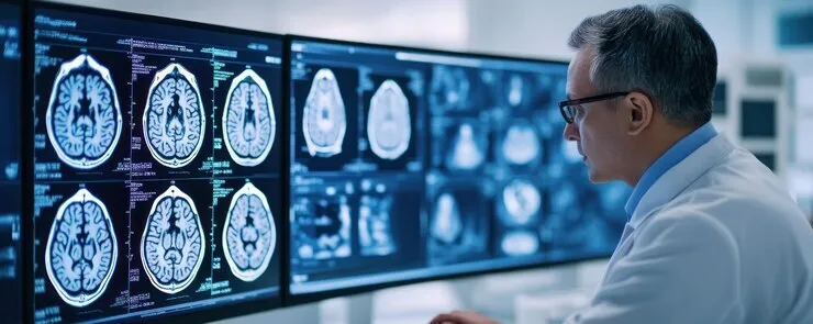
- Magnetic Resonance Imaging (MRI):Excelling in soft tissue contrast, MRI is invaluable for brain, spinal cord, joint, and abdominal organ imaging. It produces large series of images with varying tissue weighting (T1, T2, FLAIR, DWI), demanding precise slice navigation and multi parametric analysis.
- Computed Tomography (CT Scan):Known for its speed and detail in bone and lung imaging, as well as rapid assessment of acute conditions (e.g., stroke, trauma). CT scans generate volumetric data, often across multiple phases (e.g., arterial, venous), requiring advanced 3D reconstruction and density measurement tools.
- Positron Emission Tomography (PET) / PET CT / PET MRI:Primarily used in oncology, neurology, and cardiology to visualize metabolic activity rather than just anatomy. PET scans generate functional data, which is almost always fused with anatomical CT or MRI data for accurate localization of metabolic abnormalities. This fusion is critical.
- X-rays (Digital Radiography, CR/DR):The oldest and most common form of medical imaging, providing quick, foundational assessments of bone structures, chest, and abdomen. While two dimensional, digital X-rays benefit greatly from image enhancement and precise measurement tools.
A generic DICOM viewer might display these images, but it will fall short in providing the specialized tools needed for in depth diagnostic analysis unique to each modality.
Key Considerations for Choosing a Specialty Imaging Viewer
When evaluating specialty imaging solutions or a comprehensive medical image viewer that handles diverse modalities, focus on these critical aspects:
- Image Fidelity and Rendering
Quality
The viewer must accurately render images from all modalities, preserving the subtle nuances crucial for diagnosis. This includes:
- True Color and Grayscale Representation:Accurate display of varying contrast levels, especially for MRI and CT.
- High Resolution Support:Ability to handle large datasets from high resolution scanners without pixelation or lag.
- Interpolation and Smoothing:Advanced algorithms for clear rendering during zoom and pan operations.
- Modality Specific Solutions and Workflows
This is where a general viewer differentiates from a specialty solution. The viewer must intelligently adapt its toolset based on the modality loaded:
- For MRI:
- multi parametric Viewing:Simultaneous display of T1, T2, FLAIR, DWI, ADC maps, and perfusion images side by side or linked.
- Diffusion Tensor Imaging (DTI) / Tractography:Tools to visualize white matter tracts in the brain, crucial for neurosurgical planning and assessment of neurological disorders.
- Spectroscopy Analysis:Viewing and analyzing spectral data to assess metabolic changes in tissues.
- Cardiac Function Analysis:Specialized tools for cardiac MRI, including ventricular volumetric measurements and strain analysis.
- For CT Scan:
- Advanced MPR (Multi Planar Reconstruction):Precise control over oblique and curved MPRs (e.g., for vessel analysis, spinal cord mapping).
- 3D Rendering (Volume and Surface):Robust capabilities for creating interactive 3D models of organs, bones, and vasculature, essential for surgical planning, orthopedic assessments, and patient education.
- Vessel Analysis:Tools for segmenting and measuring vessels, including calculating stenosis.
- Lung Nodule Analysis:Automated or semi automated tools for tracking lung nodule size and characteristics over time.
- Bone Density Measurement:Integration with tools for quantitative bone density assessment from CT scans.
- For PET (and PET/CT/MRI Fusion):
- SUV (Standardized Uptake Value) Measurement:Accurate calculation of metabolic activity, often with automatic segmentation and normalization.
- Precise Image Fusion and Registration:The ability to accurately overlay functional PET data with anatomical CT or MRI images, ensuring perfect alignment for precise localization of lesions. This often requires robust rigid and deformable registration algorithms.
- Segmentation Tools:Automated or manual tools to segment organs and lesions for quantitative analysis.
- For X-rays (Digital Radiography/CR/DR):
- Advanced Image Enhancement Filters:Tools for sharpening, noise reduction, and contrast enhancement specific to radiography.
- Accurate Measurement Tools:Precise linear and angular measurements, especially for orthopedic applications.
- Annotation Features:Clear and easy to use annotation tools for reports and communication.
- Magnification and Pan/Zoom:Smooth navigation of large, high resolution digital X-ray images.
- For MRI:
- Performance and Speed
Regardless of modality, the viewer must handle large datasets efficiently. This means rapid loading of studies, smooth navigation through series, and instantaneous application of rendering tools, even over varying network conditions.
- Seamless Integration with Existing Systems
A top tier DICOM viewer must integrate effortlessly with your PACS, EHR, and RIS to provide comprehensive patient context and streamline workflows. This ensures that imaging data is part of the complete patient record.
- Scalability and Accessibility (Cloud Based Advantage)
For diverse modalities, especially across multiple sites or for teleradiology, a cloud based DICOM viewer offers unmatched advantages:
- Universal Access:View any specialty image from any location, on any device, via a secure web browser.
- Centralized Archive:All modality images are stored in a single, scalable cloud archive, eliminating fragmented data.
- Reduced IT Burden:No need to install and maintain specialized software on individual workstations for each modality.
- Robust Security and Compliance
Handling sensitive patient data, regardless of modality, requires unwavering commitment to security (encryption, access controls, audit trails) and adherence to regulations like HIPAA and GDPR.
- Intuitive User Experience (UI/UX)
Even with advanced features, the interface must be intuitive and easy to navigate for clinicians across different specialties. A clean design reduces learning curves and enhances productivity.
- Vendor Support and Training
For complex specialty tools, responsive vendor support and comprehensive training are crucial to maximize the software's utility and ensure smooth clinical operations.
Try Now for Anytime Anywhere DICOM Access
Try Now !The Power of a Unified Specialty Solution Scriptoware's Approach
Instead of piecing together multiple, modality specific viewers, the ideal specialty imaging solution is a single, intelligent DICOM viewer that dynamically offers the right tools for the right image.
Scriptoware's approach delivers precisely this. Our advanced medical image viewer is designed to intelligently adapt its toolset based on the specific modality being viewed. This means:
- One Platform for All Modalities:From high resolution MRI sequences and multi phase CT scans to functional PET images and standard X-rays, our viewer handles all DICOM formats with precision.
- Modality Aware Tools:When you open an MRI, the relevant cardiac analysis or neuro mapping tools are readily available. When you open a PET/CT, sophisticated fusion and SUV measurement capabilities are at your fingertips.
- Advanced Rendering:Leveraging powerful underlying technology, we ensure optimal image fidelity and performance for even the largest and most complex specialty studies.
- Cloud Based Accessibility:Being a cloud based DICOM viewer, our solution ensures that your specialists can access these advanced tools from any location, fostering collaboration and efficiency, whether they're in a dedicated radiology suite or conducting a remote consultation.
- Integrated Workflow:Seamless integration with your PACS, EHR, and RIS ensures that specialty imaging insights are always part of the comprehensive patient record, streamlining the entire diagnostic process.
By choosing Scriptoware, you're not just getting a DICOM viewer; you're getting a comprehensive, intelligent, and flexible specialty imaging solution that empowers your clinicians with the precise tools they need for accurate diagnosis across the full spectrum of medical imaging modalities.
Conclusion
The evolution of medical imaging demands more than just a general image viewer. Choosing the right viewer for MRI, CT Scan, PET, and X-rays means selecting a specialty imaging solution that understands the unique characteristics of each modality and provides the specific, advanced tools required for accurate interpretation. By investing in a comprehensive DICOM viewer that combines universal accessibility with modality specific intelligence, healthcare providers can enhance diagnostic confidence, improve workflow efficiency, and ultimately deliver higher quality, more precise patient care.
Ready to explore a versatile imaging solution that masters every modality? [Contact Scriptoware today for a personalized demonstration of our advanced DICOM viewer!]